Shedding of the primary cell wall |
In principle the cell wall in all desmids consists of three layers: an outer, amorphous layer, a primary microfibrillar layer, and a secundary microfibrillar layer (Mix 1975, Brook 1981). In general, those layers are only to be distinguished with the aid of an electron microscope. With one exception, i.e. the separation of the primary and the secondary wall as can be observed in the group of ‘real’ desmids (Family Desmidiaceae sensu stricto) shortly after cell division.
In this group of desmids (the vast majority of all desmid genera) newly formed semicells initially are only bounded by a primary cell wall layer. When the primary wall is almost fully expanded the secondary wall is laid down (between primary wall and protoplast). By the time that the development of the secundary wall is completed the primary wall is cast off, usually going hand in hand with spatial separation of the two daughter cells.
The phenomenon of shedding of the primary wall can be particularly well observed in genera characterized by entire-outlined cells (no deep incisions, no processes), like Cosmarium, Actinotaenium and Pleurotaenium.
 Image © Henk Schulp |
Cell of Pleurotaenium ehrenbergii, characterized by cylindric semicells with a swelling at the base (marking the cell sinus) and furnished with a whorl of tiny granules at the apex. The chloroplast is in the form of longitudinal, parietal bands containing many pyrenoids.
|
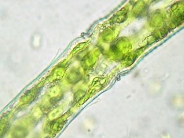 Image © Koos Meesters |
Midregion of the cell showing a ringlike istmial thickening and various chloroplast bands with globose pyrenoids. The semicell on the left side of the sinus is a young one as appears from the primary cell wall detaching the secundary wall just on the left of the basal swelling.
|
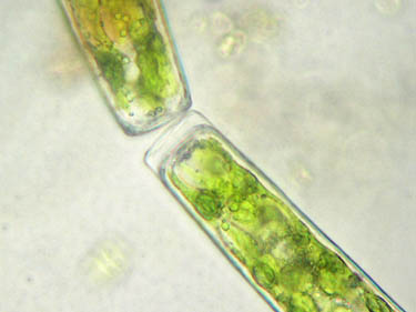 Image © Koos Meesters |
Apex of that same young semicell showing the primary cell wall detached from the secundary wall but still in contact with the primary wall of the young semicell of a sister cell.
|
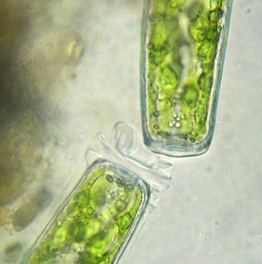 Image © Koos Meesters |
Contact region between the young semicells of two sister cells with crumpled parts of a primary wall already partly stripped off.
|
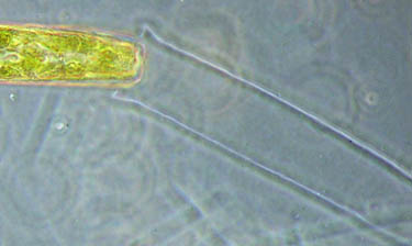 Image © Koos Meesters |
Primary wall has been skinned almost completely.
|
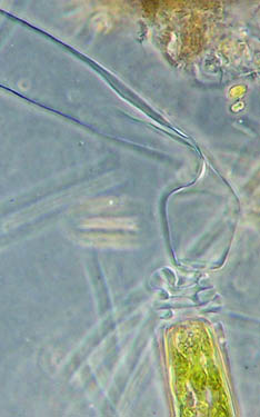 Image © Koos Meesters |
Fully detached primary wall of a young semicell still contacting the partly stripped off and crumpled primary wall of a sister cell.
|
References
Brook, A. 1981. The Biology of Desmids. — Blackwell, Oxford, 276 pp.
Mix, M., 1975 Die Feinstruktur der Zellwände der Conjugaten und ihre systematische Bedeutung. — Beihefte Nova Hedwigia 42: 179-194.