- Home
- News
- Desmidiological Communications
- Links
- Literature
- Desmid species
- A-G
- Actinotaenium
- Bambusina
- Closterium
- Cosmarium
- angulare
- amoenum
- anceps
- biretum
- botrytis
- brebissonii
- brookii
- caelatum
- cataractarum
- commissurale
- connatum
- conspersum
- cosmarioides
- crenulatum
- cyclicum
- denboeri
- dickii
- difficile
- excavatum
- fontigenum
- formosulum
- granatoides
- granatum
- hexalobum
- holmiense
- angulare
- humile
- margaritiferum
- meneghinii
- microsphinctum
- monomazum
- nasutum
- nymannianum
- obliquum
- obsoletum
- obtusatum
- ornatum
- ovale
- pachydermum
- paragranatoides
- perforatum
- pericymatium
- pokornyanum
- portianum
- protractum
- pseudoconnatum
- pseudoinsigne
- pseudopyramidatum
- punctulatum
- pyramidatum
- quinarium
- ralfsii
- regnellii
- regnesi
- reniforme
- sphyrelatum
- striolatum
- subexcavatum
- subgranatum
- subprotumidum
- taxichondriforme
- tetrachondrum
- turpinii
- ungarianum var. subtriplicatum
- venustum
- Cosmocladium
- Cylindrocystis
- Desmidium
- Docidium
- Euastrum
- Gonatozygon
- H-R
- S-X
- Sphaerozosma
- Spirotaenia
- Spondylosium
- Staurastrum
- alternans
- arachne
- bloklandiae
- brachiatum
- brebissonii
- brevispina
- chaetoceras
- cingulum
- controversum
- crassangulatum
- diacanthum
- dilatatum
- echinatum
- elongatum
- furcatum
- furcigerum
- habeebense
- heimerlianum
- hirsutum
- hystrix
- inconspicuum
- lapponicum
- levanderi
- margaritaceum
- monticulosum
- pileolatum
- pingue
- polytrichum
- punctulatum
- pyramidatum
- scabrum
- sexcostatum
- spongiosum
- teliferum
- tetracerum
- vestitum
- Staurodesmus
- Teilingia
- Tetmemorus
- Tortitaenia
- Xanthidium
- A-G
- Year
- 2024
- 2023
- December - Closterium praelongum
- November - Cosmarium microsphinctum
- October - Cosmarium pokornyanum
- September - Staurastrum inconspicuum
- August - Cosmarium crenulatum
- July - Euastrum subalpinum
- June - Staurastrum levanderi
- May - Cosmarium microsphinctum
- April - Closterium subscoticum
- March - Cosmarium impressulum
- February - Cosmarium angulare
- January - Staurastrum heimerlianum
- 2022
- December - Closterium juncidum
- November - Cosmarium nasutum
- October - Cosmarium cosmarioides
- September - Staurodesmus patens
- August - Closterium turgidum
- July - Cosmarium subexcavatum
- June - Cosmarium conspersum
- May - Cosmarium excavatum
- April - Closterium directum
- March - Cosmarium anceps
- February - Cosmarium biretum
- January - Closterium baillyanum
- 2021
- December - Staurastrum cingulum
- November - Cosmarium paragranatoides
- October - Closterium attenuatum
- September - Cosmarium granatoides
- August - Cosmarium meneghinii
- July - Cosmarium commissurale
- June - Cosmarium pseudopyramidatum
- May - Staurastrum pingue
- April - Pleurotaenium simplicissimum
- March - Cosmarium ornatum
- February - Gonatozygon aculeatum
- January - Staurastrum monticulosum
- 2020
- December - Cosmarium granatum
- November - Staurastrum tetracerum
- October - Actinotaenium cucurbitinum
- September - Staurodesmus triangularis
- August - Cosmarium regnellii
- July - Staurastrum hirsutum
- June - Euastrum ansatum
- May - Cosmarium monomazum
- April - Cosmarium regnesi
- March - Actinotaenium pinicola
- February -Staurastrum crassangulatum
- January - Euastrum biscrobiculatum
- 2019
- December - Cosmarium hexalobum
- November - Euastrum pulchellum
- October - Cosmarium sphyrelatum
- September - Cosmarium margaritiferum
- August - Xanthidium tenuissimum
- July - Euastrum denticulatum
- June - Cosmarium caelatum
- May - Cosmarium difficile
- April - Spirotaenia diplohelica
- March - Staurastrum arachne
- February - Euastrum gayanum
- January - Cosmarium nasutum
- 2018
- December - Actinotaenium kriegeri
- November - Staurastrum dilatatum
- October - Euastrum pinnatum
- September - Cosmarium pseudoconnatum
- August - Cosmarium connatum
- July - Staurastrum margaritaceum
- June -Closterium nematodes
- May - Cosmarium pachydermum
- April - Euastrum dubium
- March -Staurastrum lapponicum
- February - Cosmarium humile
- January - Actinotaenium riethii
- 2017
- December - Closterium lineatum
- November - Euastrum luetkemuelleri
- Octtober - Cosmarium pyramidatum
- September - Staurastrum punctulatum
- August - Closterium limneticum
- July - Tortitaenia bahusiensis
- June - Pleurotaenium archeri
- May - Cosmarium cyclicum
- April - Euastrum coeselii
- March - Staurastrum pileolatum
- February - Cosmarium obtusatum
- January - Closterium gracile
- 2016
- December - Staurodesmus glaber
- November - Cosmarium taxichondriforme
- October - Heimansia pusilla
- September - Pleurotaenium nodulosum
- August - Euastrum lacustre
- July - Cosmarium tetrachondrum
- June - Euastrum insulare
- May - Staurastrum brebissonii
- April - Closterium cornu
- March - Actinotaenium mooreanum
- February - Cosmarium denboeri
- January - Cosmarium cataractarum
- 2015
- December - Staurastrum pyramidatum
- November - Staurodesmus dickiei
- October - Closterium moniliferum
- September - Staurastrum controversum
- August - Euastrum ventricosum
- July - Actinotaenium inconspiquum
- June - Cosmarium formosulum
- May - Xanthidium bifidum
- April - Staurastrum brevispina
- March - Cosmarium amoenum
- February - Closterium acutum
- January - Tortitaenia trabeculata
- 2014
- December - Staurastrum echinatum
- November - Micrasterias furcata
- October - Staurastrum furcatum
- September - Cosmarium protractum
- August - Staurodesmus pterosporus
- July -Staurodesmus omearae
- June - Closterium calosporum
- May - Pleurotaenium truncatum
- April - Cosmarium portianum
- March - Sphaerozosma aubertianum
- February - Staurastrum scabrum
- January - Micrasterias radiosa
- 2013
- December - Staurodesmus dejectus
- November - Staurastrum alternans
- October - Closterium closterioides
- September - Cosmarium botrytis
- August - Euastrum pseudotuddalense
- July - Staurastrum teliferum
- June - Gonatozygon kinahanii
- May - Xanthidium variabile
- April - Actinotaenium turgidum
- March - Haplotaenium rectum
- February - Staurastrum vestitum
- January - Cosmarium obsoletum
- 2012
- December - Euastrum crassum
- November - Closterium cynthia
- October - Hyalotheca mucosa
- September - Heimansia species
- August - Actinotaenium curtum
- July - Cosmarium turpinii
- June - Staurastrum elongatum
- May - Pleurotaenium ehrenbergii
- April - Euastrum ampullaceum
- March - Closterium acerosum
- February - Roya closterioides
- January - Cosmarium quinarium
- 2011
- December - Staurastrum sexcostatum
- November - Desmidium aptogonum
- October - Actinotaenium phymatosporum
- September - Cosmarium nymannianum
- August - Xanthiddiuum basidentatum
- July - Closterium angustatum
- June - Staurastrum polytrichum
- May - Cosmarium brebissonii
- April - Actinotaenium rufescens
- March - Closterium striolatum
- February - Staurastrum bloklandiae
- January - Cosmarium subprotumidum
- 2010
- December - Pleurotaenium coronatum
- November - Actinotaenium subtile
- October - Spharozosma filiforme
- September - Docidium undulatum
- Augustus - Xanthidium fasciculatum
- July - Closterium navicula
- June - Staurastrum hystrix
- May - Cosmarium ungerianum var. subtriplicatum
- April - Euastrum insigne
- March - Cosmarium venustum
- February - Actinotaenium silvae-nigrae
- January - Penium polymorphum
- 2009
- December - Desmidium baileyi
- November - Closterium pusillum
- October - Cosmarium perforatum
- September - Gonatozygon brebissonii
- August - Cosmarium ovale
- July - Penium exiguum
- June - Cosmocladium perissum
- May - Euastrum binale
- April - Netrium oblongum
- March - Cosmarium punctulatum
- February - Tetmemorus laevis
- January - Staurodesmus cuspidatus
- 2008
- December - Penium spirostriolatum
- November - Cosmarium reniforme
- October - Docidium baculum
- September - Actinotaenium cucurbita
- August - Xanthidium octocorne
- July - Tetmemorus granulatus
- June - Cosmarium obliquum
- May - Staurastrum spongiosum
- April - Cosmarium subgranatum
- March - Xanthidium cristatum
- February - Cosmocladium constrictum
- January - Micrasterias oscitans
- 2007
- December - Cylindrocystis gracilis
- November - Micrasterias denticulata
- October - Closterium delpontei
- September - Netrium interruptum
- August - Teilingia granulata
- July - Euastrum bidentatum
- June - Staurastrum diacanthum
- May - Sphaerozosma vertebratum
- April - Spondylosium pulchellum
- March - Staurodesmus extensus
- February - Spirotaenia erythrocephala
- January - Euastrum elegans
- 2006
- December - Closterium setaceum
- November - Actinotaenium diplosporum
- October - Closterium lunula
- September - Euastrum pectinatum
- August - Haplotaenium minutum
- July - Gonatozygon monotaenium
- June - Cylindrocystis brebissonii
- May - Micrasterias jenneri
- April - Roya obtusa
- March - Euastrum humerosum
- February - Mesotaenium macrococcum
- January - Xanthidium armatum
- 2005
- December - Desmidium grevillei
- November - Cosmarium pericymatium
- October - Xanthidium antilopaeum
- September - Bambusina brebissonii
- August - Mesotaenium caldariorum
- July - Micrasterias papillifera
- June - Micrasterias rotata
- May - Pleurotaenium trabecula
- April - Cosmarium ralfsii
- March - Closterium costatum
- February - Micrasterias brachyptera
- January - Tetmemorus brebissonii
- 2004
- December - Euastrum oblongum
- November - Staurodesmus mucronatus
- October - Staurastrum furcigerum
- September - Cosmarium striolatum
- August - Tortitaenia obscura
- July - Spirotaenia condensata
- June - Cosmocladium saxonicum
- May - Micrasterias truncata
- April - Cosmarium holmiense
- March - Penium margaritaceum
- February - Micrasterias apiculata
- January - Staurastrum habeebense
- 2003
- December - Netrium digitus
- November - Micrasterias pinnatifida
- October - Closterium aciculare
- September - Spondylosium ellipticum
- August - Desmidium swartzii
- July - Micrasterias fimbriata
- June - Actinotaenium didymocarpum
- May - Hyalotheca dissiliens
- April - Cosmarium pseudoinsigne
- March - Euastrum verrucosum
- February - Staurodesmus convergens
- January - Staurastrum brachiatum
- 2002
- Additions
- Euastrum humerosum (December)
- Micrasterias oscitans (June)
- Cosmarium cyclicum (June)
- Cosmarium obliquum (March)
- 2017
- Actinotaenium mooreanum (March)
- 2016
- Rotifer eating Micrasterias rotata (September)
- Netrium digitus
- Bambusina brebissonii (June)
- Actinotaenium subtile (January)
- 2015
- Spondylosium pulchellum
- Micrasterias rotata
- Pleurotaenium trabecula - (September)
- Micrasterias crux-melitensis
- Micrasterias brachyptera
- Micrasterias apiculata (August)
- Micrasterias denticulata
- Micrasterias pinnatifida (July)
- Actinotaenium diplosporum (May)
- Cosmarium botrytis
- Sphaerozosma aubertianum - (March)
- Closterium costatum (March)
- 2014
- Sphaerozosma filiforme (November)
- Penium spirostriolatum (September)
- Actinotaenium diplosporum (August)
- Penium margaritaceum (July)
- Euastrum pectinatum (June)
- Desmidium aptogonum (May)
- Staurastrum spongiosum (April)
- Micrasterias papillifera (April)
- Euastrum insigne (April)
- Desmidium baileyi (March)
- Cosmarium ovale (February)
- Staurastrum furcigerum (February)
- Euastrum germanicum (January)
- Euastrum bidentatum (December)
- Cosmarium striolatum (November)
- Cosmarium perforatum (October)
- Desmid biology
- Contact
|
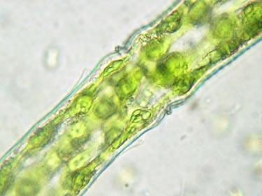
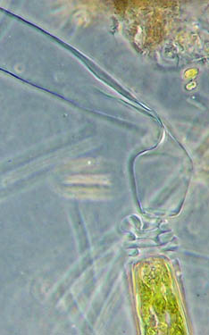
Shedding of the primary cell wallIn principle the cell wall in all desmids consists of three layers: an outer, amorphous layer, a primary microfibrillar layer, and a secundary microfibrillar layer (Mix 1975, Brook 1981). In general, those layers are only to be distinguished with the aid of an electron microscope. With one exception, i.e. the separation of the primary and the secondary wall as can be observed in the group of ‘real’ desmids (Family Desmidiaceae sensu stricto) shortly after cell division. In this group of desmids (the vast majority of all desmid genera) newly formed semicells initially are only bounded by a primary cell wall layer. When the primary wall is almost fully expanded the secondary wall is laid down (between primary wall and protoplast). By the time that the development of the secundary wall is completed the primary wall is cast off, usually going hand in hand with spatial separation of the two daughter cells. The phenomenon of shedding of the primary wall can be particularly well observed in genera characterized by entire-outlined cells (no deep incisions, no processes), like Cosmarium, Actinotaenium and Pleurotaenium.
|
Shedding of primary wall in Pleurotaenium ehrenbergii |
|
|
Cell of Pleurotaenium ehrenbergii, characterized by cylindric semicells with a swelling at the base (marking the cell sinus) and furnished with a whorl of tiny granules at the apex. The chloroplast is in the form of longitudinal, parietal bands containing many pyrenoids.
|
|
Midregion of the cell showing a ringlike istmial thickening and various chloroplast bands with globose pyrenoids. The semicell on the left side of the sinus is a young one as appears from the primary cell wall detaching the secundary wall just on the left of the basal swelling.
|
|
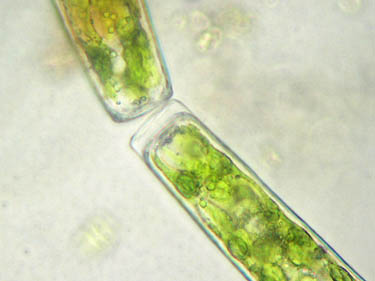
Apex of that same young semicell showing the primary cell wall detached from the secundary wall but still in contact with the primary wall of the young semicell of a sister cell.
|
|
Contact region between the young semicells of two sister cells with crumpled parts of a primary wall already partly stripped off.
|
|
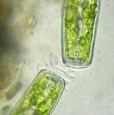
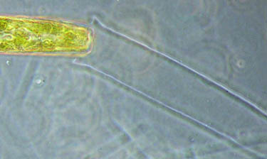
Primary wall has been skinned almost completely.
|
|
Fully detached primary wall of a young semicell still contacting the partly stripped off and crumpled primary wall of a sister cell.
|
References Brook, A. 1981. The Biology of Desmids. — Blackwell, Oxford, 276 pp. Mix, M., 1975 Die Feinstruktur der Zellwände der Conjugaten und ihre systematische Bedeutung. — Beihefte Nova Hedwigia 42: 179-194. |
|