Desmids and fungal parasites


Image © Alfred van Geest

Cell of Staurastrum rotula (in apical view) set with sporangia of a chytrid fungus.


Image © Koos Meesters

Cell of Closterium littorale on its back bearing an encysted chytrid (containing a lipid globule).

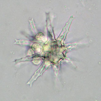
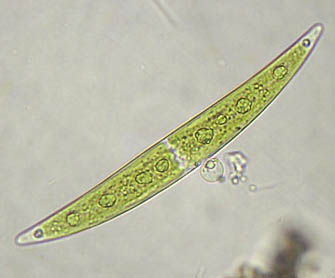
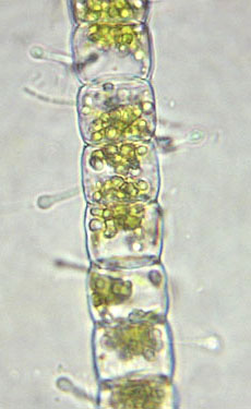
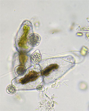
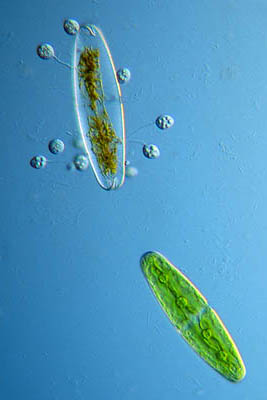
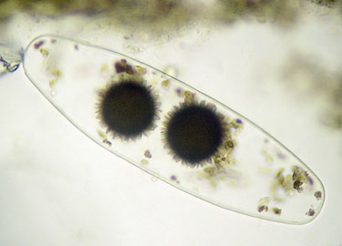
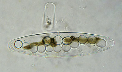
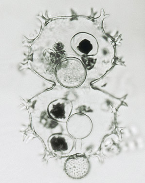
Desmids not only are threatened by predators (like amoebes) and grazers (like water-fleas) but also by parasitic fungi (see Teilingia ). Some species are extremely sensitive to fungal infection, such as the tropical species Staurastrum rotula, complete populations of which often appear to be infested by a chytrid fungus.

Identification of fungal parasites usually is very difficult as they but rarely show species-specific morphological structures. Sometimes, however, the relationship between host and parasite is  such specific that identification of the host implies identification of the fungal parasite.

Aquatic fungi reproduce by means of flagellate spores (zoospores). Based on the number of flagella per spore a commonly made difference is that between monoflagellate fungi and biflagellate fungi (Canter-Lund & Lund, 1995).

The thallus (plant body) of a monoflagellate fungus (or chytrid) is most simple, i.e., a single protoplast that, after a period of growth, becomes a sporangium and divides up into zoospores. If a motile, naked zoospore meets a susceptible algal cell it may attach to the cell wall, encyst and subsequently produce a fine germ tube that penetrates the algal wall. Enzymes produced by the chytrid break down the content of the algal cell which is then transferred back into the encysted spore. As a result, the latter enlarges and eventually in its turn becomes a sporangium. For that matter, infection by a single chytrid not necessarily needs to lead to the death of the algal host cell.


Image © Koos Meesters

Filament of Hyalotheca dissiliens infected by some chytrid fungus (genus Zygorhizidium?)


Image © Koos Meesters

Sporangia of some chytrid (Zygorhizidium?) on cells of Euastrum ansatum


Image © Wim van Egmond (mouseover)

Two cells of Tetmemorus laevis, the left one being set with chytrids.


A special group of chytrid fungi is specifically found only in conjugate algae.  After a zoospore has encysted on an algal cell, its contents pass into the cell. The naked, amoeboid fungal body then enlarges and becomes thick-walled. The content of this body is released to form a ball-shaped sorus that segments into a number of sporangia. The monoflagellate zoospores escape  through a rupture in the killed algal host cell.


Image © Koos Meesters ( mouseover )

Two thick-walled, spiny bodies of the chytrid genus Micromyces parasitizing a cell of Netrium digitus.


In contrast to monoflagellate chytrids, the body of a biflagellate fungus that grows inside an algal cell may develop a (large) number of sporangia each opening to the outer world by way of an exit tube.


Cell of Netrium digitus killed by a biflagellate fungus of the genus Mizocytium. Notice the many globular sporangia, each  provided with an exit tube to release their spores.

Image © Koos Meesters


Cell of Xanthidium armatum infected by a biflagellate fungus of the genus Micromycopsis. In the empty algal cell are several so-called prosori. Each endobiotic prosorus forms an exit tube to release a globular, exobiotic sorus that develops a thick, granulate wall. When mature, it opens to release several sporangia which, in their turn burst to release the biflagellate spores.

Image © IBED


Reference:

Canter-Lund, H. & J.W.G. Lund. 1995. Freshwater Algae — their microscopic world explored. Biopress, Bristol.