- Home
- News
- Reports
- Desmidiological Communications
- Links
- Literature
- Desmid species
- A-G
- Actinotaenium
- Bambusina
- Closterium
- acerosum
- aciculare
- acutum
- angustatum
- attenuatum
- baillyanum
- calosporum
- closterioides
- cornu
- costatum
- cynthia
- delpontei
- dianae
- directum
- gracile
- incurvum
- juncidum
- karnakense
- kuetzingii
- limneticum
- lineatum
- lunula
- moniliferum
- navicula
- nematodes
- praelongum
- pusillum
- ralfsii
- rostratum
- setaceum
- striolatum
- submoniliferum
- subscoticum
- tortitaenioides
- turgidum
- Cosmarium
- angulare
- amoenum
- anceps
- beckii
- benedictum
- biretum
- botrytis
- brebissonii
- brookii
- caelatum
- cataractarum
- commissurale
- connatum
- conspersum
- cosmarioides
- crenatoides
- crenulatum
- cyclicum
- debaryi
- denboeri
- depressum
- dickii
- difficile
- dilatatum
- excavatum
- fontigenum
- formosulum
- granatoides
- granatum
- hexalobum
- holmiense
- hornavanense
- hostensiense
- angulare
- humile
- margaritiferum
- meneghinii
- microsphinctum
- monomazum
- nasutum
- neocrenatum
- notabile
- nymannianum
- obliquum
- obsoletum
- obtusatum
- ornatum
- ovale
- pachydermum
- paragranatoides
- perforatum
- pericymatium
- pokornyanum
- portianum
- protractum
- pseudoconnatum
- pseudoinsigne
- pseudopyramidatum
- punctulatum
- pseudoprotuberans
- pyramidatum
- quadratum
- quinarium
- ralfsii
- regnellii
- regnesi
- reniforme
- June - Cosmarium rostafinskii
- scenedesmus
- sphyrelatum
- striolatum
- subcostatulum
- subexcavatum
- subgranatum
- subprotumidum
- subpunctulatum
- taxichondriforme
- tetrachondrum
- tetraophthalmum
- turpinii
- ungarianum var. subtriplicatum
- venustum
- Cosmocladium
- Cylindrocystis
- Desmidium
- Docidium
- Euastrum
- Gonatozygon
- H-R
- S-X
- Sphaerozosma
- Spirotaenia
- Spondylosium
- Staurastrum
- alternans
- anatinum
- arachne
- bloklandiae
- brachiatum
- brebissonii
- brevispina
- chaetoceras
- cingulum
- controversum
- crassangulatum
- diacanthum
- dilatatum
- echinatum
- elongatum
- furcatum
- furcigerum
- groenbladii
- habeebense
- heimerlianum
- hirsutum
- hystrix
- inconspicuum
- kouwetsii
- lapponicum
- levanderii
- manfeldtii
- margaritaceum
- monticulosum
- paradoxum
- pileolatum
- pingue
- polytrichum
- punctulatum
- pyramidatum
- scabrum
- sexcostatum
- spongiosum
- striolatum
- teliferum
- tetracerum
- vestitum
- Staurodesmus
- Teilingia
- Tetmemorus
- Tortitaenia
- Xanthidium
- A-G
- Year
- 2026
- 2025
- December - Staurastrum paradoxum
- November - Cosmarium tetraophthalmum
- October - Closterium ralfsii
- September - Cosmarium hornavanense
- August - Closterium dianae
- July - Staurastrum anatinum
- June - Cosmarium rostafinskii
- May - Staurastrum groenbladii
- April - Cosmarium beckii
- March - Cosmarium subcostatulum
- February - Closterium rostratum
- January - Cosmarium pseudoprotuberans
- 2024
- December - Cosmarium neocrenatum
- November - Closterium submoniliferum
- October - Cosmarium hostensiense
- September - Closterium kuetzingii
- August - Cosmarium scenedesmus
- July - Cosmarium quadratum
- June - Euastrum minimum
- May - Staurastrum manfeldtii
- April - Closterium incurvum
- March - Cosmarium fontigenum
- February - Cosmarium dickii
- January - Euastrum didelta
- 2023
- December - Closterium praelongum
- November - Cosmarium microsphinctum
- October - Cosmarium pokornyanum
- September - Staurastrum inconspicuum
- August - Cosmarium crenulatum
- July - Euastrum subalpinum
- June - Staurastrum levanderi
- May - Cosmarium microsphinctum
- April - Closterium subscoticum
- March - Cosmarium impressulum
- February - Cosmarium angulare
- January - Staurastrum heimerlianum
- 2022
- December - Closterium juncidum
- November - Cosmarium nasutum
- October - Cosmarium cosmarioides
- September - Staurodesmus patens
- August - Closterium turgidum
- July - Cosmarium subexcavatum
- June - Cosmarium conspersum
- May - Cosmarium excavatum
- April - Closterium directum
- March - Cosmarium anceps
- February - Cosmarium biretum
- January - Closterium baillyanum
- 2021
- December - Staurastrum cingulum
- November - Cosmarium paragranatoides
- October - Closterium attenuatum
- September - Cosmarium granatoides
- August - Cosmarium meneghinii
- July - Cosmarium commissurale
- June - Cosmarium pseudopyramidatum
- May - Staurastrum pingue
- April - Pleurotaenium simplicissimum
- March - Cosmarium ornatum
- February - Gonatozygon aculeatum
- January - Staurastrum monticulosum
- 2020
- December - Cosmarium granatum
- November - Staurastrum tetracerum
- October - Actinotaenium cucurbitinum
- September - Staurodesmus triangularis
- August - Cosmarium regnellii
- July - Staurastrum hirsutum
- June - Euastrum ansatum
- May - Cosmarium monomazum
- April - Cosmarium regnesi
- March - Actinotaenium pinicola
- February -Staurastrum crassangulatum
- January - Euastrum biscrobiculatum
- 2019
- December - Cosmarium hexalobum
- November - Euastrum pulchellum
- October - Cosmarium sphyrelatum
- September - Cosmarium margaritiferum
- August - Xanthidium tenuissimum
- July - Euastrum denticulatum
- June - Cosmarium caelatum
- May - Cosmarium difficile
- April - Spirotaenia diplohelica
- March - Staurastrum arachne
- February - Euastrum gayanum
- January - Cosmarium nasutum
- 2018
- December - Actinotaenium kriegeri
- November - Staurastrum dilatatum
- October - Euastrum pinnatum
- September - Cosmarium pseudoconnatum
- August - Cosmarium connatum
- July - Staurastrum margaritaceum
- June -Closterium nematodes
- May - Cosmarium pachydermum
- April - Euastrum dubium
- March -Staurastrum lapponicum
- February - Cosmarium humile
- January - Actinotaenium riethii
- 2017
- December - Closterium lineatum
- November - Euastrum luetkemuelleri
- Octtober - Cosmarium pyramidatum
- September - Staurastrum punctulatum
- August - Closterium limneticum
- July - Tortitaenia bahusiensis
- June - Pleurotaenium archeri
- May - Cosmarium cyclicum
- April - Euastrum coeselii
- March - Staurastrum pileolatum
- February - Cosmarium obtusatum
- January - Closterium gracile
- 2016
- December - Staurodesmus glaber
- November - Cosmarium taxichondriforme
- October - Heimansia pusilla
- September - Pleurotaenium nodulosum
- August - Euastrum lacustre
- July - Cosmarium tetrachondrum
- June - Euastrum insulare
- May - Staurastrum brebissonii
- April - Closterium cornu
- March - Actinotaenium mooreanum
- February - Cosmarium denboeri
- January - Cosmarium cataractarum
- 2015
- December - Staurastrum pyramidatum
- November - Staurodesmus dickiei
- October - Closterium moniliferum
- September - Staurastrum controversum
- August - Euastrum ventricosum
- July - Actinotaenium inconspiquum
- June - Cosmarium formosulum
- May - Xanthidium bifidum
- April - Staurastrum brevispina
- March - Cosmarium amoenum
- February - Closterium acutum
- January - Tortitaenia trabeculata
- 2014
- December - Staurastrum echinatum
- November - Micrasterias furcata
- October - Staurastrum furcatum
- September - Cosmarium protractum
- August - Staurodesmus pterosporus
- July -Staurodesmus omearae
- June - Closterium calosporum
- May - Pleurotaenium truncatum
- April - Cosmarium portianum
- March - Sphaerozosma aubertianum
- February - Staurastrum scabrum
- January - Micrasterias radiosa
- 2013
- December - Staurodesmus dejectus
- November - Staurastrum alternans
- October - Closterium closterioides
- September - Cosmarium botrytis
- August - Euastrum pseudotuddalense
- July - Staurastrum teliferum
- June - Gonatozygon kinahanii
- May - Xanthidium variabile
- April - Actinotaenium turgidum
- March - Haplotaenium rectum
- February - Staurastrum vestitum
- January - Cosmarium obsoletum
- 2012
- December - Euastrum crassum
- November - Closterium cynthia
- October - Hyalotheca mucosa
- September - Heimansia species
- August - Actinotaenium curtum
- July - Cosmarium turpinii
- June - Staurastrum elongatum
- May - Pleurotaenium ehrenbergii
- April - Euastrum ampullaceum
- March - Closterium acerosum
- February - Roya closterioides
- January - Cosmarium quinarium
- 2011
- December - Staurastrum sexcostatum
- November - Desmidium aptogonum
- October - Actinotaenium phymatosporum
- September - Cosmarium nymannianum
- August - Xanthiddiuum basidentatum
- July - Closterium angustatum
- June - Staurastrum polytrichum
- May - Cosmarium brebissonii
- April - Actinotaenium rufescens
- March - Closterium striolatum
- February - Staurastrum bloklandiae
- January - Cosmarium subprotumidum
- 2010
- December - Pleurotaenium coronatum
- November - Actinotaenium subtile
- October - Spharozosma filiforme
- September - Docidium undulatum
- Augustus - Xanthidium fasciculatum
- July - Closterium navicula
- June - Staurastrum hystrix
- May - Cosmarium ungerianum var. subtriplicatum
- April - Euastrum insigne
- March - Cosmarium venustum
- February - Actinotaenium silvae-nigrae
- January - Penium polymorphum
- 2009
- December - Desmidium baileyi
- November - Closterium pusillum
- October - Cosmarium perforatum
- September - Gonatozygon brebissonii
- August - Cosmarium ovale
- July - Penium exiguum
- June - Cosmocladium perissum
- May - Euastrum binale
- April - Netrium oblongum
- March - Cosmarium subpunctulatum
- February - Tetmemorus laevis
- January - Staurodesmus cuspidatus
- 2008
- December - Penium spirostriolatum
- November - Cosmarium reniforme
- October - Docidium baculum
- September - Actinotaenium cucurbita
- August - Xanthidium octocorne
- July - Tetmemorus granulatus
- June - Cosmarium obliquum
- May - Staurastrum spongiosum
- April - Cosmarium subgranatum
- March - Xanthidium cristatum
- February - Cosmocladium constrictum
- January - Micrasterias oscitans
- 2007
- December - Cylindrocystis gracilis
- November - Micrasterias denticulata
- October - Closterium delpontei
- September - Netrium interruptum
- August - Teilingia granulata
- July - Euastrum bidentatum
- June - Staurastrum diacanthum
- May - Sphaerozosma vertebratum
- April - Spondylosium pulchellum
- March - Staurodesmus extensus
- February - Spirotaenia erythrocephala
- January - Euastrum elegans
- 2006
- December - Closterium setaceum
- November - Actinotaenium diplosporum
- October - Closterium lunula
- September - Euastrum pectinatum
- August - Haplotaenium minutum
- July - Gonatozygon monotaenium
- June - Cylindrocystis brebissonii
- May - Micrasterias jenneri
- April - Roya obtusa
- March - Euastrum humerosum
- February - Mesotaenium macrococcum
- January - Xanthidium armatum
- 2005
- December - Desmidium grevillei
- November - Cosmarium pericymatium
- October - Xanthidium antilopaeum
- September - Bambusina brebissonii
- August - Mesotaenium caldariorum
- July - Micrasterias papillifera
- June - Micrasterias rotata
- May - Pleurotaenium trabecula
- April - Cosmarium ralfsii
- March - Closterium costatum
- February - Micrasterias brachyptera
- January - Tetmemorus brebissonii
- 2004
- December - Euastrum oblongum
- November - Staurodesmus mucronatus
- October - Staurastrum furcigerum
- September - Cosmarium striolatum
- August - Tortitaenia obscura
- July - Spirotaenia condensata
- June - Cosmocladium saxonicum
- May - Micrasterias truncata
- April - Cosmarium holmiense
- March - Penium margaritaceum
- February - Micrasterias apiculata
- January - Staurastrum habeebense
- 2003
- December - Netrium digitus
- November - Micrasterias pinnatifida
- October - Closterium aciculare
- September - Spondylosium ellipticum
- August - Desmidium swartzii
- July - Micrasterias fimbriata
- June - Actinotaenium didymocarpum
- May - Hyalotheca dissiliens
- April - Cosmarium pseudoinsigne
- March - Euastrum verrucosum
- February - Staurodesmus convergens
- January - Staurastrum brachiatum
- 2002
- Additions
- 2017
- Actinotaenium mooreanum (March)
- 2016
- Rotifer eating Micrasterias rotata (September)
- Netrium digitus
- Bambusina brebissonii (June)
- Actinotaenium subtile (January)
- 2015
- Spondylosium pulchellum
- Micrasterias rotata
- Pleurotaenium trabecula - (September)
- Micrasterias crux-melitensis
- Micrasterias brachyptera
- Micrasterias apiculata (August)
- Micrasterias denticulata
- Micrasterias pinnatifida (July)
- Actinotaenium diplosporum (May)
- Cosmarium botrytis
- Sphaerozosma aubertianum - (March)
- Closterium costatum (March)
- 2014
- Sphaerozosma filiforme (November)
- Penium spirostriolatum (September)
- Actinotaenium diplosporum (August)
- Penium margaritaceum (July)
- Euastrum pectinatum (June)
- Desmidium aptogonum (May)
- Staurastrum spongiosum (April)
- Micrasterias papillifera (April)
- Euastrum insigne (April)
- Desmidium baileyi (March)
- Cosmarium ovale (February)
- Staurastrum furcigerum (February)
- Euastrum germanicum (January)
- Euastrum bidentatum (December)
- Cosmarium striolatum (November)
- Cosmarium perforatum (October)
- Biology
- Contact
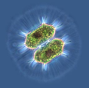
| Mucilaginous cell envelope |
Cell of Xanthidium fasciculatum enveloped by a distinct mucilaginous layer. The outside of the envelope is marked by numerous bar-shaped micro-particles (presumably bacteria). |
|
| In
many desmid species cells are surrounded by a thick, sharply outlined gelatinous
layer. The mucous cell envelope usually encloses the complete cell body inclusive
of its possible processes. The occurrence of sharply bordered cell envelopes
is taxon-linked. We don’t meet it in, e.g., the genera Closterium and Micrasterias whereas it is a rather common phenomenon in, e.g., the genera Cosmarium, Staurastrum and Staurodesmus. Though, also within
those latter genera there are species lacking a distinct envelope.
The function of the extracellular mucous envelope is not quite clear. Because density of the mucous is lower than that of the proper cell in planktic species it will reduce sinking velocity. As an envelope increases particle size considerably, it also could obstruct grazing by zooplankton. A third hypothesis is that it might help in trapping scarce, dissolved nutrients. Arguing for that latter hypothesis is that desmid species characteristic of eutrophic (nutrient-rich) habitats are in want of a distinct envelope (Coesel 1994). Reference: Coesel, P.F.M., 1994. On the ecological significance of a cellular mucilaginous envelope in planktic desmids. — Algological Studies 73: 65-74. |
||
 |
Cells of Staurodesmus cuspidatus var. curvatus in Indian ink suspension (not penetrating into the mucous envelope).
Image © IBED |
Cells of the oligo-mesotrophic taxon Cosmarium abbreviatum var. planctonicum (with envelope) and the eutrophic species Staurastrum chaetoceras (without envelope) in a mixed continuous flow culture showing that the occurrence of an extracellular envelope is genotypically, not phenotypically determined. Image © IBED |
 |
|
Cell of Xanthidium antilopaeum (in phase contrast) showing a radiate, fibrillar structure of the envelope.
|
![]()