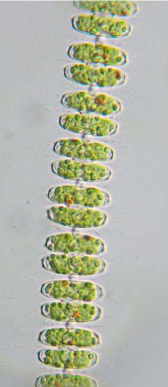
Image © Koos Meesters
Filament of Sphaerozosma aubertianum. Notice ellipsoid semicells, on their lateral sides marked by granule-like mucous extrusions.
Cell dimensions (L x B): ca 20 x 20 µm
- Home
- Desmid species
- A-G
- Actinotaenium
- Bambusina
- Closterium
- Cosmarium
- angulare
- amoenum
- anceps
- biretum
- botrytis
- brebissonii
- caelatum
- cataractarum
- commissurale
- connatum
- conspersum
- cosmarioides
- cyclicum
- denboeri
- difficile
- excavatum
- formosulum
- granatoides
- granatum
- hexalobum
- holmiense
- humile
- impressulum
- margaritiferum
- meneghinii
- microsphinctum
- monomazum
- nasutum
- nymannianum
- obliquum
- obsoletum
- obtusatum
- ornatum
- ovale
- paragranatoides
- perforatum
- pericymatium
- portianum
- protractum
- pseudoconnatum
- pseudoinsigne
- pseudopyramidatum
- punctulatum
- pyramidatum
- quinarium
- ralfsii
- regnellii
- regnesi
- reniforme
- sphyrelatum
- striolatum
- subexcavatum
- subgranatum
- subprotumidum
- taxichondriforme
- tetrachondrum
- turpinii
- ungarianum var. subtriplicatum
- venustum
- Cosmocladium
- Cylindrocystis
- Desmidium
- Docidium
- Euastrum
- Gonatozygon
- H-R
- S-X
- Sphaerozosma
- Spirotaenia
- Spondylosium
- Staurastrum
- alternans
- arachne
- bloklandiae
- brachiatum
- brebissonii
- brevispina
- chaetoceras
- cingulum
- controversum
- crassangulatum
- diacanthum
- dilatatum
- echinatum
- elongatum
- furcatum
- furcigerum
- habeebense
- heimerlianum
- hirsutum
- hystrix
- lapponicum
- levanderi
- margaritaceum
- monticulosum
- pileolatum
- pingue
- polytrichum
- punctulatum
- pyramidatum
- scabrum
- sexcostatum
- spongiosum
- teliferum
- tetracerum
- vestitum
- Staurodesmus
- Teilingia
- Tetmemorus
- Tortitaenia
- Xanthidium
- A-G
- Year
- 2023
- 2022
- December - Closterium juncidum
- November - Cosmarium nasutum
- October - Cosmarium cosmarioides
- September - Staurodesmus patens
- August - Closterium turgidum
- July - Cosmarium subexcavatum
- June - Cosmarium conspersum
- May - Cosmarium excavatum
- April - Closterium directum
- March - Cosmarium anceps
- February - Cosmarium biretum
- January - Closterium baillyanum
- 2021
- December - Staurastrum cingulum
- November - Cosmarium paragranatoides
- October - Closterium attenuatum
- September - Cosmarium granatoides
- August - Cosmarium meneghinii
- July - Cosmarium commissurale
- June - Cosmarium pseudopyramidatum
- May - Staurastrum pingue
- April - Pleurotaenium simplicissimum
- March - Cosmarium ornatum
- February - Gonatozygon aculeatum
- January - Staurastrum monticulosum
- 2020
- December - Cosmarium granatum
- November - Staurastrum tetracerum
- October - Actinotaenium cucurbitinum
- September - Staurodesmus triangularis
- August - Cosmarium regnellii
- July - Staurastrum hirsutum
- June - Euastrum ansatum
- May - Cosmarium monomazum
- April - Cosmarium regnesi
- March - Actinotaenium pinicola
- February -Staurastrum crassangulatum
- January - Euastrum biscrobiculatum
- 2019
- December - Cosmarium hexalobum
- November - Euastrum pulchellum
- October - Cosmarium sphyrelatum
- September - Cosmarium margaritiferum
- August - Xanthidium tenuissimum
- July - Euastrum denticulatum
- June - Cosmarium caelatum
- May - Cosmarium difficile
- April - Spirotaenia diplohelica
- March - Staurastrum arachne
- February - Euastrum gayanum
- January - Cosmarium nasutum
- 2018
- December - Actinotaenium kriegeri
- November - Staurastrum dilatatum
- October - Euastrum pinnatum
- September - Cosmarium pseudoconnatum
- August - Cosmarium connatum
- July - Staurastrum margaritaceum
- June -Closterium nematodes
- May - Cosmarium pachydermum
- April - Euastrum dubium
- March -Staurastrum lapponicum
- February - Cosmarium humile
- January - Actinotaenium riethii
- 2017
- December - Closterium lineatum
- November - Euastrum luetkemuelleri
- Octtober - Cosmarium pyramidatum
- September - Staurastrum punctulatum
- August - Closterium limneticum
- July - Tortitaenia bahusiensis
- June - Pleurotaenium archeri
- May - Cosmarium cyclicum
- April - Euastrum coeselii
- March - Staurastrum pileolatum
- February - Cosmarium obtusatum
- January - Closterium gracile
- 2016
- December - Staurodesmus glaber
- November - Cosmarium taxichondriforme
- October - Heimansia pusilla
- September - Pleurotaenium nodulosum
- August - Euastrum lacustre
- July - Cosmarium tetrachondrum
- June - Euastrum insulare
- May - Staurastrum brebissonii
- April - Closterium cornu
- March - Actinotaenium mooreanum
- February - Cosmarium denboeri
- January - Cosmarium cataractarum
- 2015
- December - Staurastrum pyramidatum
- November - Staurodesmus dickiei
- October - Closterium moniliferum
- September - Staurastrum controversum
- August - Euastrum ventricosum
- July - Actinotaenium inconspiquum
- June - Cosmarium formosulum
- May - Xanthidium bifidum
- April - Staurastrum brevispina
- March - Cosmarium amoenum
- February - Closterium acutum
- January - Tortitaenia trabeculata
- 2014
- December - Staurastrum echinatum
- November - Micrasterias furcata
- October - Staurastrum furcatum
- September - Cosmarium protractum
- August - Staurodesmus pterosporus
- July -Staurodesmus omearae
- June - Closterium calosporum
- May - Pleurotaenium truncatum
- April - Cosmarium portianum
- March - Sphaerozosma aubertianum
- February - Staurastrum scabrum
- January - Micrasterias radiosa
- 2013
- December - Staurodesmus dejectus
- November - Staurastrum alternans
- October - Closterium closterioides
- September - Cosmarium botrytis
- August - Euastrum pseudotuddalense
- July - Staurastrum teliferum
- June - Gonatozygon kinahanii
- May - Xanthidium variabile
- April - Actinotaenium turgidum
- March - Haplotaenium rectum
- February - Staurastrum vestitum
- January - Cosmarium obsoletum
- 2012
- December - Euastrum crassum
- November - Closterium cynthia
- October - Hyalotheca mucosa
- September - Heimansia species
- August - Actinotaenium curtum
- July - Cosmarium turpinii
- June - Staurastrum elongatum
- May - Pleurotaenium ehrenbergii
- April - Euastrum ampullaceum
- March - Closterium acerosum
- February - Roya closterioides
- January - Cosmarium quinarium
- 2011
- December - Staurastrum sexcostatum
- November - Desmidium aptogonum
- October - Actinotaenium phymatosporum
- September - Cosmarium nymannianum
- August - Xanthiddiuum basidentatum
- July - Closterium angustatum
- June - Staurastrum polytrichum
- May - Cosmarium brebissonii
- April - Actinotaenium rufescens
- March - Closterium striolatum
- February - Staurastrum bloklandiae
- January - Cosmarium subprotumidum
- 2010
- December - Pleurotaenium coronatum
- November - Actinotaenium subtile
- October - Spharozosma filiforme
- September - Docidium undulatum
- Augustus - Xanthidium fasciculatum
- July - Closterium navicula
- June - Staurastrum hystrix
- May - Cosmarium ungerianum var. subtriplicatum
- April - Euastrum insigne
- March - Cosmarium venustum
- February - Actinotaenium silvae-nigrae
- January - Penium polymorphum
- 2009
- December - Desmidium baileyi
- November - Closterium pusillum
- October - Cosmarium perforatum
- September - Gonatozygon brebissonii
- August - Cosmarium ovale
- July - Penium exiguum
- June - Cosmocladium perissum
- May - Euastrum binale
- April - Netrium oblongum
- March - Cosmarium punctulatum
- February - Tetmemorus laevis
- January - Staurodesmus cuspidatus
- 2008
- December - Penium spirostriolatum
- November - Cosmarium reniforme
- October - Docidium baculum
- September - Actinotaenium cucurbita
- August - Xanthidium octocorne
- July - Tetmemorus granulatus
- June - Cosmarium obliquum
- May - Staurastrum spongiosum
- April - Cosmarium subgranatum
- March - Xanthidium cristatum
- February - Cosmocladium constrictum
- January - Micrasterias oscitans
- 2007
- December - Cylindrocystis gracilis
- November - Micrasterias denticulata
- October - Closterium delpontei
- September - Netrium interruptum
- August - Teilingia granulata
- July - Euastrum bidentatum
- June - Staurastrum diacanthum
- May - Sphaerozosma vertebratum
- April - Spondylosium pulchellum
- March - Staurodesmus extensus
- February - Spirotaenia erythrocephala
- January - Euastrum elegans
- 2006
- December - Closterium setaceum
- November - Actinotaenium diplosporum
- October - Closterium lunula
- September - Euastrum pectinatum
- August - Haplotaenium minutum
- July - Gonatozygon monotaenium
- June - Cylindrocystis brebissonii
- May - Micrasterias jenneri
- April - Roya obtusa
- March - Euastrum humerosum
- February - Mesotaenium macrococcum
- January - Xanthidium armatum
- 2005
- December - Desmidium grevillei
- November - Cosmarium pericymatium
- October - Xanthidium antilopaeum
- September - Bambusina brebissonii
- August - Mesotaenium caldariorum
- July - Micrasterias papillifera
- June - Micrasterias rotata
- May - Pleurotaenium trabecula
- April - Cosmarium ralfsii
- March - Closterium costatum
- February - Micrasterias brachyptera
- January - Tetmemorus brebissonii
- 2004
- December - Euastrum oblongum
- November - Staurodesmus mucronatus
- October - Staurastrum furcigerum
- September - Cosmarium striolatum
- August - Tortitaenia obscura
- July - Spirotaenia condensata
- June - Cosmocladium saxonicum
- May - Micrasterias truncata
- April - Cosmarium holmiense
- March - Penium margaritaceum
- February - Micrasterias apiculata
- January - Staurastrum habeebense
- 2003
- December - Netrium digitus
- November - Micrasterias pinnatifida
- October - Closterium aciculare
- September - Spondylosium ellipticum
- August - Desmidium swartzii
- July - Micrasterias fimbriata
- June - Actinotaenium didymocarpum
- May - Hyalotheca dissiliens
- April - Cosmarium pseudoinsigne
- March - Euastrum verrucosum
- February - Staurodesmus convergens
- January - Staurastrum brachiatum
- 2002
- Additions
- Euastrum humerosum (December)
- Micrasterias oscitans (June)
- Cosmarium cyclicum (June)
- Cosmarium obliquum (March)
- 2017
- Actinotaenium mooreanum (March)
- 2016
- Rotifer eating Micrasterias rotata (September)
- Netrium digitus
- Bambusina brebissonii (June)
- Actinotaenium subtile (January)
- 2015
- Spondylosium pulchellum
- Micrasterias rotata
- Pleurotaenium trabecula - (September)
- Micrasterias crux-melitensis
- Micrasterias brachyptera
- Micrasterias apiculata (August)
- Micrasterias denticulata
- Micrasterias pinnatifida (July)
- Actinotaenium diplosporum (May)
- Cosmarium botrytis
- Sphaerozosma aubertianum - (March)
- Closterium costatum (March)
- 2014
- Sphaerozosma filiforme (November)
- Penium spirostriolatum (September)
- Actinotaenium diplosporum (August)
- Penium margaritaceum (July)
- Euastrum pectinatum (June)
- Desmidium aptogonum (May)
- Staurastrum spongiosum (April)
- Micrasterias papillifera (April)
- Euastrum insigne (April)
- Desmidium baileyi (March)
- Cosmarium ovale (February)
- Staurastrum furcigerum (February)
- Euastrum germanicum (January)
- Euastrum bidentatum (December)
- Cosmarium striolatum (November)
- Cosmarium perforatum (October)
Desmid of the month
March 2014
Sphaerozosma aubertianum
Sphaerozosma aubertianum very much resembles Sph. vertebratum. Differences between those two species are subtle and, maybe, partly questionable (Coesel & Van Westen 2013). Sph. aubertianum is said to differ from Sph. vertebratum by more pronounced cell wall pores, marked by granule-like gelatinous extrusions. Moreover, semicell shape is elliptic rather than rounded-oblong, resulting in a wider opened cell sinus. Whereas the globose zygospore in Sph. aubertianum is furnished with long spines, that in Sph. vertebratum (only recorded in a few nineteenth century publications) is described as smooth-walled. In the Netherlands, Sph. aubertianum is a rare species, recently encountered in some mesotrophic moorland pools.
Reference:
Coesel, P.F.M. & Van Westen, M.C., 2013. Taxonomic notes on Dutch desmids V (Streptophyta, Desmidiales): new species, new morphological features. — Phytotaxa 84: 46-54.
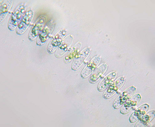 Image © Koos Meesters
Filament of Sph. aubertianum with (almost) empty cells. Notice transversal rows of cell wall pores: two in each semicell.
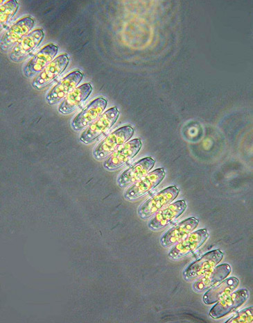 Image © Koos Meesters
Sph. aubertianum fotographed with interference contrast showing a filament that is surrounded by a thick mucilaginous sheath.
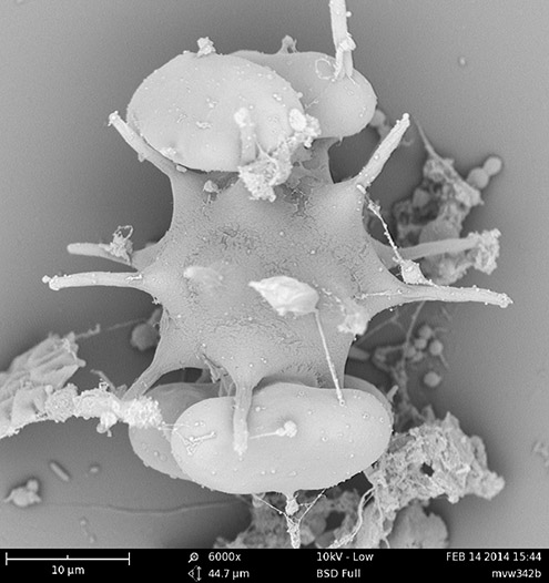
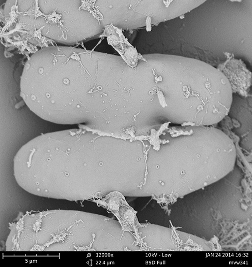 SEM image © Marien van Westen
Cell of Sph. aubertianum, SEM image.
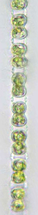
Image © Koos Meesters
Filament of Sph. aubertianum in lateral view better showing the interconnecting apical cell processes.
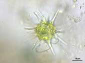
Image © Marien van Westen
Spiny zygospore of Sph. aubertianum.
 SEM image © Marien van Westen
Zygospore of Sph. aubertianum, SEM image.