|
Desmid of the month
February 2010
Actinotaenium silvae-nigrae
Actinotaenium silvae-nigrae (in most floras listed as Penium silvae-nigrae) is often confused with Penium polymorphum. The two species are of similar shape and size whereas their cell wall shows a pattern of longitudinal striae. Ultrastructural cell wall studies, however, indicated that the present species of the month is marked by a pore type that is characteristic of the family Desmidiaceae, not that of the Peniaceae (Kouwets & Coesel 1984). Additional differences between Actinotaenium silvae nigrae and Penium polymorphum are in the shape of the chloroplast, the occurrence of girdle bands and the nature of the longitudinal cell wall striation. In the Netherlands, A. silvae-nigrae is of occasional occurrence in acidic, oligotrophic bog pools.
Reference:
Kouwets, F.A.C. & Coesel, P.F.M., 1984. Taxonomic revision of the conjugatophycean family Peniacae on the basis of cell wall ultrastructure. — Journal of Phycology 20: 555- 562.
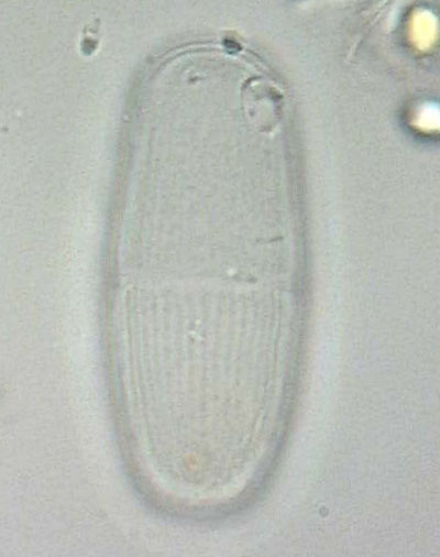
Image © Marien van Westen
Empty cell of A. silvae-nigrae. Notice that longitudinal cell wall striation is more faint and less fine than in P. polymorphum.
Images Hanny Kooijman-van Blokland © IBED
|
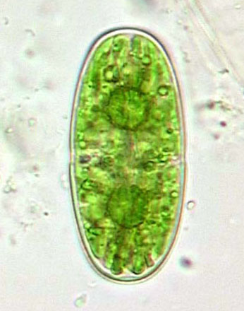
Image © Marien van Westen
Cell of Actinotaenium silvae-nigrae. As compared to Penium polymorphum, chloroplast ribbons are less regular in shape. Girdle bands do not occur.
Cell dimensions (L x B): ca 60 x 25 µm
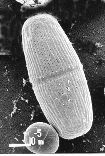
Image Hanny Kooijman-van Blokland © IBED
SEM picture of A. silvae-nigrae. Notice that cell wall striation is in the form of longitudinal plications, not as sharply marked ridges like in P. polymorphum.
<<<
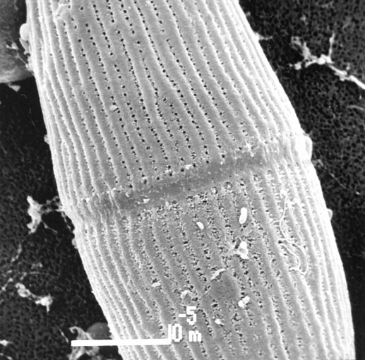
Detail of midregion of the cell. Cell wall pores are located in longitudinal rows in between the cell wall plications.
mouse over:
Detail of cell apex. Mucilage extrusions by the cell wall pores render them a somewhat granulate appearance. Notice that there are no distinct cell wall plications in this picture suggesting that those plications could be an artefact caused by fixation. |