 Image
©
Henk Schulp
Image
©
Henk Schulp
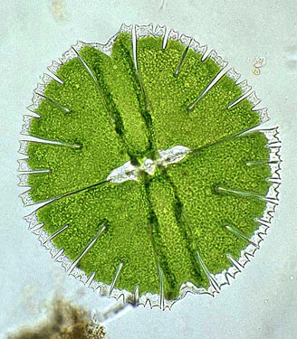
Cell of Micrasterias rotata. Note the gradual widening of the median incision of the apical lobe and the emarginate lobules at the cell margin.
Cell dimensions (L x B): ca 280 x 250 µm
- Home
- Desmid species
- A-G
- Actinotaenium
- Bambusina
- Closterium
- Cosmarium
- angulare
- amoenum
- anceps
- biretum
- botrytis
- brebissonii
- caelatum
- cataractarum
- commissurale
- connatum
- conspersum
- cosmarioides
- cyclicum
- denboeri
- difficile
- excavatum
- formosulum
- granatoides
- granatum
- hexalobum
- holmiense
- humile
- impressulum
- margaritiferum
- meneghinii
- microsphinctum
- monomazum
- nasutum
- nymannianum
- obliquum
- obsoletum
- obtusatum
- ornatum
- ovale
- paragranatoides
- perforatum
- pericymatium
- portianum
- protractum
- pseudoconnatum
- pseudoinsigne
- pseudopyramidatum
- punctulatum
- pyramidatum
- quinarium
- ralfsii
- regnellii
- regnesi
- reniforme
- sphyrelatum
- striolatum
- subexcavatum
- subgranatum
- subprotumidum
- taxichondriforme
- tetrachondrum
- turpinii
- ungarianum var. subtriplicatum
- venustum
- Cosmocladium
- Cylindrocystis
- Desmidium
- Docidium
- Euastrum
- Gonatozygon
- H-R
- S-X
- Sphaerozosma
- Spirotaenia
- Spondylosium
- Staurastrum
- alternans
- arachne
- bloklandiae
- brachiatum
- brebissonii
- brevispina
- chaetoceras
- cingulum
- controversum
- crassangulatum
- diacanthum
- dilatatum
- echinatum
- elongatum
- furcatum
- furcigerum
- habeebense
- heimerlianum
- hirsutum
- hystrix
- lapponicum
- levanderi
- margaritaceum
- monticulosum
- pileolatum
- pingue
- polytrichum
- punctulatum
- pyramidatum
- scabrum
- sexcostatum
- spongiosum
- teliferum
- tetracerum
- vestitum
- Staurodesmus
- Teilingia
- Tetmemorus
- Tortitaenia
- Xanthidium
- A-G
- Year
- 2023
- 2022
- December - Closterium juncidum
- November - Cosmarium nasutum
- October - Cosmarium cosmarioides
- September - Staurodesmus patens
- August - Closterium turgidum
- July - Cosmarium subexcavatum
- June - Cosmarium conspersum
- May - Cosmarium excavatum
- April - Closterium directum
- March - Cosmarium anceps
- February - Cosmarium biretum
- January - Closterium baillyanum
- 2021
- December - Staurastrum cingulum
- November - Cosmarium paragranatoides
- October - Closterium attenuatum
- September - Cosmarium granatoides
- August - Cosmarium meneghinii
- July - Cosmarium commissurale
- June - Cosmarium pseudopyramidatum
- May - Staurastrum pingue
- April - Pleurotaenium simplicissimum
- March - Cosmarium ornatum
- February - Gonatozygon aculeatum
- January - Staurastrum monticulosum
- 2020
- December - Cosmarium granatum
- November - Staurastrum tetracerum
- October - Actinotaenium cucurbitinum
- September - Staurodesmus triangularis
- August - Cosmarium regnellii
- July - Staurastrum hirsutum
- June - Euastrum ansatum
- May - Cosmarium monomazum
- April - Cosmarium regnesi
- March - Actinotaenium pinicola
- February -Staurastrum crassangulatum
- January - Euastrum biscrobiculatum
- 2019
- December - Cosmarium hexalobum
- November - Euastrum pulchellum
- October - Cosmarium sphyrelatum
- September - Cosmarium margaritiferum
- August - Xanthidium tenuissimum
- July - Euastrum denticulatum
- June - Cosmarium caelatum
- May - Cosmarium difficile
- April - Spirotaenia diplohelica
- March - Staurastrum arachne
- February - Euastrum gayanum
- January - Cosmarium nasutum
- 2018
- December - Actinotaenium kriegeri
- November - Staurastrum dilatatum
- October - Euastrum pinnatum
- September - Cosmarium pseudoconnatum
- August - Cosmarium connatum
- July - Staurastrum margaritaceum
- June -Closterium nematodes
- May - Cosmarium pachydermum
- April - Euastrum dubium
- March -Staurastrum lapponicum
- February - Cosmarium humile
- January - Actinotaenium riethii
- 2017
- December - Closterium lineatum
- November - Euastrum luetkemuelleri
- Octtober - Cosmarium pyramidatum
- September - Staurastrum punctulatum
- August - Closterium limneticum
- July - Tortitaenia bahusiensis
- June - Pleurotaenium archeri
- May - Cosmarium cyclicum
- April - Euastrum coeselii
- March - Staurastrum pileolatum
- February - Cosmarium obtusatum
- January - Closterium gracile
- 2016
- December - Staurodesmus glaber
- November - Cosmarium taxichondriforme
- October - Heimansia pusilla
- September - Pleurotaenium nodulosum
- August - Euastrum lacustre
- July - Cosmarium tetrachondrum
- June - Euastrum insulare
- May - Staurastrum brebissonii
- April - Closterium cornu
- March - Actinotaenium mooreanum
- February - Cosmarium denboeri
- January - Cosmarium cataractarum
- 2015
- December - Staurastrum pyramidatum
- November - Staurodesmus dickiei
- October - Closterium moniliferum
- September - Staurastrum controversum
- August - Euastrum ventricosum
- July - Actinotaenium inconspiquum
- June - Cosmarium formosulum
- May - Xanthidium bifidum
- April - Staurastrum brevispina
- March - Cosmarium amoenum
- February - Closterium acutum
- January - Tortitaenia trabeculata
- 2014
- December - Staurastrum echinatum
- November - Micrasterias furcata
- October - Staurastrum furcatum
- September - Cosmarium protractum
- August - Staurodesmus pterosporus
- July -Staurodesmus omearae
- June - Closterium calosporum
- May - Pleurotaenium truncatum
- April - Cosmarium portianum
- March - Sphaerozosma aubertianum
- February - Staurastrum scabrum
- January - Micrasterias radiosa
- 2013
- December - Staurodesmus dejectus
- November - Staurastrum alternans
- October - Closterium closterioides
- September - Cosmarium botrytis
- August - Euastrum pseudotuddalense
- July - Staurastrum teliferum
- June - Gonatozygon kinahanii
- May - Xanthidium variabile
- April - Actinotaenium turgidum
- March - Haplotaenium rectum
- February - Staurastrum vestitum
- January - Cosmarium obsoletum
- 2012
- December - Euastrum crassum
- November - Closterium cynthia
- October - Hyalotheca mucosa
- September - Heimansia species
- August - Actinotaenium curtum
- July - Cosmarium turpinii
- June - Staurastrum elongatum
- May - Pleurotaenium ehrenbergii
- April - Euastrum ampullaceum
- March - Closterium acerosum
- February - Roya closterioides
- January - Cosmarium quinarium
- 2011
- December - Staurastrum sexcostatum
- November - Desmidium aptogonum
- October - Actinotaenium phymatosporum
- September - Cosmarium nymannianum
- August - Xanthiddiuum basidentatum
- July - Closterium angustatum
- June - Staurastrum polytrichum
- May - Cosmarium brebissonii
- April - Actinotaenium rufescens
- March - Closterium striolatum
- February - Staurastrum bloklandiae
- January - Cosmarium subprotumidum
- 2010
- December - Pleurotaenium coronatum
- November - Actinotaenium subtile
- October - Spharozosma filiforme
- September - Docidium undulatum
- Augustus - Xanthidium fasciculatum
- July - Closterium navicula
- June - Staurastrum hystrix
- May - Cosmarium ungerianum var. subtriplicatum
- April - Euastrum insigne
- March - Cosmarium venustum
- February - Actinotaenium silvae-nigrae
- January - Penium polymorphum
- 2009
- December - Desmidium baileyi
- November - Closterium pusillum
- October - Cosmarium perforatum
- September - Gonatozygon brebissonii
- August - Cosmarium ovale
- July - Penium exiguum
- June - Cosmocladium perissum
- May - Euastrum binale
- April - Netrium oblongum
- March - Cosmarium punctulatum
- February - Tetmemorus laevis
- January - Staurodesmus cuspidatus
- 2008
- December - Penium spirostriolatum
- November - Cosmarium reniforme
- October - Docidium baculum
- September - Actinotaenium cucurbita
- August - Xanthidium octocorne
- July - Tetmemorus granulatus
- June - Cosmarium obliquum
- May - Staurastrum spongiosum
- April - Cosmarium subgranatum
- March - Xanthidium cristatum
- February - Cosmocladium constrictum
- January - Micrasterias oscitans
- 2007
- December - Cylindrocystis gracilis
- November - Micrasterias denticulata
- October - Closterium delpontei
- September - Netrium interruptum
- August - Teilingia granulata
- July - Euastrum bidentatum
- June - Staurastrum diacanthum
- May - Sphaerozosma vertebratum
- April - Spondylosium pulchellum
- March - Staurodesmus extensus
- February - Spirotaenia erythrocephala
- January - Euastrum elegans
- 2006
- December - Closterium setaceum
- November - Actinotaenium diplosporum
- October - Closterium lunula
- September - Euastrum pectinatum
- August - Haplotaenium minutum
- July - Gonatozygon monotaenium
- June - Cylindrocystis brebissonii
- May - Micrasterias jenneri
- April - Roya obtusa
- March - Euastrum humerosum
- February - Mesotaenium macrococcum
- January - Xanthidium armatum
- 2005
- December - Desmidium grevillei
- November - Cosmarium pericymatium
- October - Xanthidium antilopaeum
- September - Bambusina brebissonii
- August - Mesotaenium caldariorum
- July - Micrasterias papillifera
- June - Micrasterias rotata
- May - Pleurotaenium trabecula
- April - Cosmarium ralfsii
- March - Closterium costatum
- February - Micrasterias brachyptera
- January - Tetmemorus brebissonii
- 2004
- December - Euastrum oblongum
- November - Staurodesmus mucronatus
- October - Staurastrum furcigerum
- September - Cosmarium striolatum
- August - Tortitaenia obscura
- July - Spirotaenia condensata
- June - Cosmocladium saxonicum
- May - Micrasterias truncata
- April - Cosmarium holmiense
- March - Penium margaritaceum
- February - Micrasterias apiculata
- January - Staurastrum habeebense
- 2003
- December - Netrium digitus
- November - Micrasterias pinnatifida
- October - Closterium aciculare
- September - Spondylosium ellipticum
- August - Desmidium swartzii
- July - Micrasterias fimbriata
- June - Actinotaenium didymocarpum
- May - Hyalotheca dissiliens
- April - Cosmarium pseudoinsigne
- March - Euastrum verrucosum
- February - Staurodesmus convergens
- January - Staurastrum brachiatum
- 2002
- Additions
- Euastrum humerosum (December)
- Micrasterias oscitans (June)
- Cosmarium cyclicum (June)
- Cosmarium obliquum (March)
- 2017
- Actinotaenium mooreanum (March)
- 2016
- Rotifer eating Micrasterias rotata (September)
- Netrium digitus
- Bambusina brebissonii (June)
- Actinotaenium subtile (January)
- 2015
- Spondylosium pulchellum
- Micrasterias rotata
- Pleurotaenium trabecula - (September)
- Micrasterias crux-melitensis
- Micrasterias brachyptera
- Micrasterias apiculata (August)
- Micrasterias denticulata
- Micrasterias pinnatifida (July)
- Actinotaenium diplosporum (May)
- Cosmarium botrytis
- Sphaerozosma aubertianum - (March)
- Closterium costatum (March)
- 2014
- Sphaerozosma filiforme (November)
- Penium spirostriolatum (September)
- Actinotaenium diplosporum (August)
- Penium margaritaceum (July)
- Euastrum pectinatum (June)
- Desmidium aptogonum (May)
- Staurastrum spongiosum (April)
- Micrasterias papillifera (April)
- Euastrum insigne (April)
- Desmidium baileyi (March)
- Cosmarium ovale (February)
- Staurastrum furcigerum (February)
- Euastrum germanicum (January)
- Euastrum bidentatum (December)
- Cosmarium striolatum (November)
- Cosmarium perforatum (October)
Desmid of the month
June 2005
Micrasterias rotata
M. rotata may be confused with some other Micrasterias species, in particular M. thomasiana and M. fimbriata. From M. thomasiana it differs in the shape of the apical median incision (relatively narrow and clear-cut in M. thomasiana, gradually widening in M. rotata), from M. fimbriata in the shape of the highest-order lobules (spinous-denticulate in M. fimbriata, emarginate in M. rotata).
Although M. rotata holds as one of the commoner Micrasterias species, in the Netherlands its distribution is seriously curtailed due to deterioration of the natural habitat. Today M. rotata is only occasionally encountered in mesotrophic moorland pools, peat pits and fen hollows.
Image
© Henk Schulp
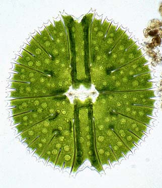
Another cell of M. rotata showing two prominent, longitudinal chloroplast ridges in each of the apical lobes as well as many scattered globose pyrenoids.
 Image © André Vanhoof
Spiny zygospore of Micrasterias rotata.
 Image © André Vanhoof
Detail of zygospore showing single spines some of which being hook-shaped at the end.
see also
"The
process of asexual reproduction" showing a dividing
Micrasterias rotata.