 image © Marien van Westen
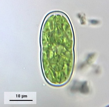
Actinotaenium phymatosporum. Note the two asteroid-stelloid chloroplasts each enclosing a globular pyrenoid.
Cell dimensions (L x B): 33 x 15 µm
- Home
- Desmid species
- A-G
- Actinotaenium
- Bambusina
- Closterium
- Cosmarium
- angulare
- amoenum
- anceps
- biretum
- botrytis
- brebissonii
- caelatum
- cataractarum
- commissurale
- connatum
- conspersum
- cosmarioides
- cyclicum
- denboeri
- difficile
- excavatum
- formosulum
- granatoides
- granatum
- hexalobum
- holmiense
- humile
- impressulum
- margaritiferum
- meneghinii
- microsphinctum
- monomazum
- nasutum
- nymannianum
- obliquum
- obsoletum
- obtusatum
- ornatum
- ovale
- paragranatoides
- perforatum
- pericymatium
- portianum
- protractum
- pseudoconnatum
- pseudoinsigne
- pseudopyramidatum
- punctulatum
- pyramidatum
- quinarium
- ralfsii
- regnellii
- regnesi
- reniforme
- sphyrelatum
- striolatum
- subexcavatum
- subgranatum
- subprotumidum
- taxichondriforme
- tetrachondrum
- turpinii
- ungarianum var. subtriplicatum
- venustum
- Cosmocladium
- Cylindrocystis
- Desmidium
- Docidium
- Euastrum
- Gonatozygon
- H-R
- S-X
- Sphaerozosma
- Spirotaenia
- Spondylosium
- Staurastrum
- alternans
- arachne
- bloklandiae
- brachiatum
- brebissonii
- brevispina
- chaetoceras
- cingulum
- controversum
- crassangulatum
- diacanthum
- dilatatum
- echinatum
- elongatum
- furcatum
- furcigerum
- habeebense
- heimerlianum
- hirsutum
- hystrix
- lapponicum
- levanderi
- margaritaceum
- monticulosum
- pileolatum
- pingue
- polytrichum
- punctulatum
- pyramidatum
- scabrum
- sexcostatum
- spongiosum
- teliferum
- tetracerum
- vestitum
- Staurodesmus
- Teilingia
- Tetmemorus
- Tortitaenia
- Xanthidium
- A-G
- Year
- 2023
- 2022
- December - Closterium juncidum
- November - Cosmarium nasutum
- October - Cosmarium cosmarioides
- September - Staurodesmus patens
- August - Closterium turgidum
- July - Cosmarium subexcavatum
- June - Cosmarium conspersum
- May - Cosmarium excavatum
- April - Closterium directum
- March - Cosmarium anceps
- February - Cosmarium biretum
- January - Closterium baillyanum
- 2021
- December - Staurastrum cingulum
- November - Cosmarium paragranatoides
- October - Closterium attenuatum
- September - Cosmarium granatoides
- August - Cosmarium meneghinii
- July - Cosmarium commissurale
- June - Cosmarium pseudopyramidatum
- May - Staurastrum pingue
- April - Pleurotaenium simplicissimum
- March - Cosmarium ornatum
- February - Gonatozygon aculeatum
- January - Staurastrum monticulosum
- 2020
- December - Cosmarium granatum
- November - Staurastrum tetracerum
- October - Actinotaenium cucurbitinum
- September - Staurodesmus triangularis
- August - Cosmarium regnellii
- July - Staurastrum hirsutum
- June - Euastrum ansatum
- May - Cosmarium monomazum
- April - Cosmarium regnesi
- March - Actinotaenium pinicola
- February -Staurastrum crassangulatum
- January - Euastrum biscrobiculatum
- 2019
- December - Cosmarium hexalobum
- November - Euastrum pulchellum
- October - Cosmarium sphyrelatum
- September - Cosmarium margaritiferum
- August - Xanthidium tenuissimum
- July - Euastrum denticulatum
- June - Cosmarium caelatum
- May - Cosmarium difficile
- April - Spirotaenia diplohelica
- March - Staurastrum arachne
- February - Euastrum gayanum
- January - Cosmarium nasutum
- 2018
- December - Actinotaenium kriegeri
- November - Staurastrum dilatatum
- October - Euastrum pinnatum
- September - Cosmarium pseudoconnatum
- August - Cosmarium connatum
- July - Staurastrum margaritaceum
- June -Closterium nematodes
- May - Cosmarium pachydermum
- April - Euastrum dubium
- March -Staurastrum lapponicum
- February - Cosmarium humile
- January - Actinotaenium riethii
- 2017
- December - Closterium lineatum
- November - Euastrum luetkemuelleri
- Octtober - Cosmarium pyramidatum
- September - Staurastrum punctulatum
- August - Closterium limneticum
- July - Tortitaenia bahusiensis
- June - Pleurotaenium archeri
- May - Cosmarium cyclicum
- April - Euastrum coeselii
- March - Staurastrum pileolatum
- February - Cosmarium obtusatum
- January - Closterium gracile
- 2016
- December - Staurodesmus glaber
- November - Cosmarium taxichondriforme
- October - Heimansia pusilla
- September - Pleurotaenium nodulosum
- August - Euastrum lacustre
- July - Cosmarium tetrachondrum
- June - Euastrum insulare
- May - Staurastrum brebissonii
- April - Closterium cornu
- March - Actinotaenium mooreanum
- February - Cosmarium denboeri
- January - Cosmarium cataractarum
- 2015
- December - Staurastrum pyramidatum
- November - Staurodesmus dickiei
- October - Closterium moniliferum
- September - Staurastrum controversum
- August - Euastrum ventricosum
- July - Actinotaenium inconspiquum
- June - Cosmarium formosulum
- May - Xanthidium bifidum
- April - Staurastrum brevispina
- March - Cosmarium amoenum
- February - Closterium acutum
- January - Tortitaenia trabeculata
- 2014
- December - Staurastrum echinatum
- November - Micrasterias furcata
- October - Staurastrum furcatum
- September - Cosmarium protractum
- August - Staurodesmus pterosporus
- July -Staurodesmus omearae
- June - Closterium calosporum
- May - Pleurotaenium truncatum
- April - Cosmarium portianum
- March - Sphaerozosma aubertianum
- February - Staurastrum scabrum
- January - Micrasterias radiosa
- 2013
- December - Staurodesmus dejectus
- November - Staurastrum alternans
- October - Closterium closterioides
- September - Cosmarium botrytis
- August - Euastrum pseudotuddalense
- July - Staurastrum teliferum
- June - Gonatozygon kinahanii
- May - Xanthidium variabile
- April - Actinotaenium turgidum
- March - Haplotaenium rectum
- February - Staurastrum vestitum
- January - Cosmarium obsoletum
- 2012
- December - Euastrum crassum
- November - Closterium cynthia
- October - Hyalotheca mucosa
- September - Heimansia species
- August - Actinotaenium curtum
- July - Cosmarium turpinii
- June - Staurastrum elongatum
- May - Pleurotaenium ehrenbergii
- April - Euastrum ampullaceum
- March - Closterium acerosum
- February - Roya closterioides
- January - Cosmarium quinarium
- 2011
- December - Staurastrum sexcostatum
- November - Desmidium aptogonum
- October - Actinotaenium phymatosporum
- September - Cosmarium nymannianum
- August - Xanthiddiuum basidentatum
- July - Closterium angustatum
- June - Staurastrum polytrichum
- May - Cosmarium brebissonii
- April - Actinotaenium rufescens
- March - Closterium striolatum
- February - Staurastrum bloklandiae
- January - Cosmarium subprotumidum
- 2010
- December - Pleurotaenium coronatum
- November - Actinotaenium subtile
- October - Spharozosma filiforme
- September - Docidium undulatum
- Augustus - Xanthidium fasciculatum
- July - Closterium navicula
- June - Staurastrum hystrix
- May - Cosmarium ungerianum var. subtriplicatum
- April - Euastrum insigne
- March - Cosmarium venustum
- February - Actinotaenium silvae-nigrae
- January - Penium polymorphum
- 2009
- December - Desmidium baileyi
- November - Closterium pusillum
- October - Cosmarium perforatum
- September - Gonatozygon brebissonii
- August - Cosmarium ovale
- July - Penium exiguum
- June - Cosmocladium perissum
- May - Euastrum binale
- April - Netrium oblongum
- March - Cosmarium punctulatum
- February - Tetmemorus laevis
- January - Staurodesmus cuspidatus
- 2008
- December - Penium spirostriolatum
- November - Cosmarium reniforme
- October - Docidium baculum
- September - Actinotaenium cucurbita
- August - Xanthidium octocorne
- July - Tetmemorus granulatus
- June - Cosmarium obliquum
- May - Staurastrum spongiosum
- April - Cosmarium subgranatum
- March - Xanthidium cristatum
- February - Cosmocladium constrictum
- January - Micrasterias oscitans
- 2007
- December - Cylindrocystis gracilis
- November - Micrasterias denticulata
- October - Closterium delpontei
- September - Netrium interruptum
- August - Teilingia granulata
- July - Euastrum bidentatum
- June - Staurastrum diacanthum
- May - Sphaerozosma vertebratum
- April - Spondylosium pulchellum
- March - Staurodesmus extensus
- February - Spirotaenia erythrocephala
- January - Euastrum elegans
- 2006
- December - Closterium setaceum
- November - Actinotaenium diplosporum
- October - Closterium lunula
- September - Euastrum pectinatum
- August - Haplotaenium minutum
- July - Gonatozygon monotaenium
- June - Cylindrocystis brebissonii
- May - Micrasterias jenneri
- April - Roya obtusa
- March - Euastrum humerosum
- February - Mesotaenium macrococcum
- January - Xanthidium armatum
- 2005
- December - Desmidium grevillei
- November - Cosmarium pericymatium
- October - Xanthidium antilopaeum
- September - Bambusina brebissonii
- August - Mesotaenium caldariorum
- July - Micrasterias papillifera
- June - Micrasterias rotata
- May - Pleurotaenium trabecula
- April - Cosmarium ralfsii
- March - Closterium costatum
- February - Micrasterias brachyptera
- January - Tetmemorus brebissonii
- 2004
- December - Euastrum oblongum
- November - Staurodesmus mucronatus
- October - Staurastrum furcigerum
- September - Cosmarium striolatum
- August - Tortitaenia obscura
- July - Spirotaenia condensata
- June - Cosmocladium saxonicum
- May - Micrasterias truncata
- April - Cosmarium holmiense
- March - Penium margaritaceum
- February - Micrasterias apiculata
- January - Staurastrum habeebense
- 2003
- December - Netrium digitus
- November - Micrasterias pinnatifida
- October - Closterium aciculare
- September - Spondylosium ellipticum
- August - Desmidium swartzii
- July - Micrasterias fimbriata
- June - Actinotaenium didymocarpum
- May - Hyalotheca dissiliens
- April - Cosmarium pseudoinsigne
- March - Euastrum verrucosum
- February - Staurodesmus convergens
- January - Staurastrum brachiatum
- 2002
- Additions
- Euastrum humerosum (December)
- Micrasterias oscitans (June)
- Cosmarium cyclicum (June)
- Cosmarium obliquum (March)
- 2017
- Actinotaenium mooreanum (March)
- 2016
- Rotifer eating Micrasterias rotata (September)
- Netrium digitus
- Bambusina brebissonii (June)
- Actinotaenium subtile (January)
- 2015
- Spondylosium pulchellum
- Micrasterias rotata
- Pleurotaenium trabecula - (September)
- Micrasterias crux-melitensis
- Micrasterias brachyptera
- Micrasterias apiculata (August)
- Micrasterias denticulata
- Micrasterias pinnatifida (July)
- Actinotaenium diplosporum (May)
- Cosmarium botrytis
- Sphaerozosma aubertianum - (March)
- Closterium costatum (March)
- 2014
- Sphaerozosma filiforme (November)
- Penium spirostriolatum (September)
- Actinotaenium diplosporum (August)
- Penium margaritaceum (July)
- Euastrum pectinatum (June)
- Desmidium aptogonum (May)
- Staurastrum spongiosum (April)
- Micrasterias papillifera (April)
- Euastrum insigne (April)
- Desmidium baileyi (March)
- Cosmarium ovale (February)
- Staurastrum furcigerum (February)
- Euastrum germanicum (January)
- Euastrum bidentatum (December)
- Cosmarium striolatum (November)
- Cosmarium perforatum (October)
Desmid of the month
October 2011
Actinotaenium phymatosporum
Actinotaenium phymatosporum (in most floras listed as Penium phymatosporum) may be readily confused with Actinotaenium spinospermum (Penium spinospermum). Both species are characterized by small cell dimensions and a faintly striate cell wall (often the striae are hardly visible). Actually, the striae are made of longitudinal series of closely set cell wall pores, better to be distinguished as the pores are marked by protruding excretion products. Owing to the nature of this cell wall sculpturing both above-mentioned species were transferred from Penium to Actinotaenium (Kouwets & Coesel 1984). Vegetative cells in A. phymatosporum are slightly longer than in A. spinospermum but the main difference is in the shape of the zygospore: irregularly rectangular in A. phymatosporum versus about globose in A. spinospermum. In the Netherlands, A. phymatosporum is known from acidic, oligo-mesotrophic, shallow water bodies. Zygospores are but rarely encountered.
 image © Marien van Westen
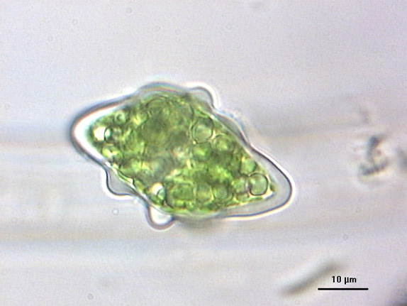
Dead, empty cell of A. phymatosporum. Notice faint striation of the cell wall.
Reference:
Kouwets, F.A.C. & Coesel, P.F.M., 1984. Taxonomic revision of the conjugatophycean family Peniacae on the basis of cell wall ultrastructure. — Journal of Phycology 20: 555- 562.
 mouse-over images Hanny Kooijman – van Blokland, © IBED
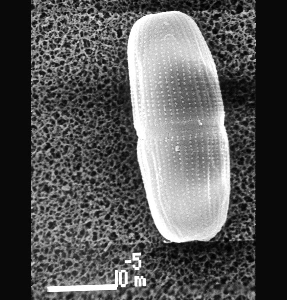
SEM picture of A. phymatosporum. Notice longitudinal series of cell wall pores.
Mouse over: Detail of apex showing the piling of excreted material around the majority of the cell wall pores.
 mouse-over images © Marien van Westen
Zygospore of A. phymatosporum. Notice irregularly shape with rounded, conical protuberances.