 Image © Henk Schulp
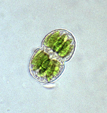
Cell of Cosmarium pericymatium characterized by a shallow sinus, semi-elliptic semicells with an unevenly undulate margin and stelloid chloroplasts.
Cell dimensions (L x B): 40 x 25 µm
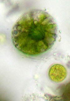
Foto © Alfred van Geest
Apical view of a C. pericymatium cell showing a broadly elliptic outline and a stelloid chloroplast.
mouse over images © Alfred van Geest
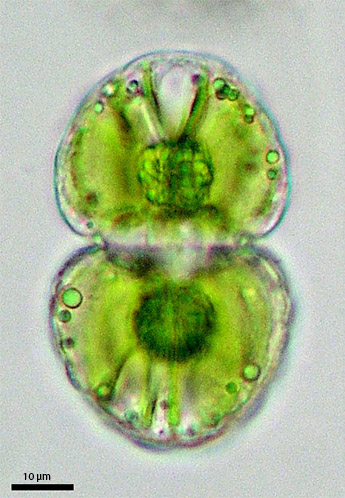
Cell of C. pericymatium (var. corrugatum) in frontal view and lateral view respectively.
* In 2023 Cosmarium pericymatium var. corrugatum was renamed Cosmarium brookii. Reference: Coesel, P.F.M. & Meesters, J., 2023. Desmids of the Lowlands, 2nd edition. KNNV Publishing, 424 pp.
Desmid of the month
November 2005
Cosmarium pericymatium*
Cosmarium pericymatium belongs to a small group of desmid species that can stand long periods of severe drought (Brook 2001). With its thick-set cell shape (low cell surface to content ratio) and thick cell wall evaporation rate is reduced. Cells are broadly elliptic to almost circular in apical view. At first glance they not seldom seem to be smooth-walled and may remind of some species of the genus Actinotaenium, but at closer examination the semi-elliptic semicells appear to be faintly and unevenly undulate at the margin. The degree of cell wall sculpturing is variable. Usually, in the apical part of the semicell a number of flattened tubercles may be distinguished whereas the basal part shows a supraisthmial ring of longitudinal plications. Depending on cell size, cell outline and markedness of cell wall sculpturing a number of varieties is distinguished the taxonomic value of which, however, is debatable.
C. pericymatium is known from wet moss cushions and shallow rain puddles, but also from garden ornaments periodically holding some water. In the Netherlands it was only rather recently discovered for the first time, i.e., amongst mosses growing on a sandstone statue located in Blijdorp Zoo (Rotterdam).
Reference
Brook, A.J., 2001. The drought-resistant desmid, Cosmarium pericymatium Nordstedt, and a description of the new var. corrugatum. — Quekett Journal of Microscopy 39: 127-132.
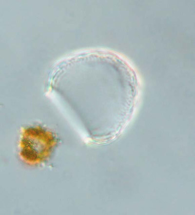
mouse over images © Koos Meesters
Empty semicell of C. pericymatium (var. corrugatum)* showing scattered, flattened tubercles in apical part of the semicell and supraisthmial whorl of longitudinal plications at the base.
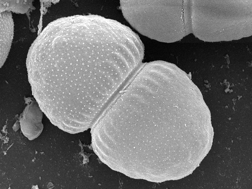 Image © Jan Šťastný (after Czech material)
SEM picture of C. pericymatium (var. corrugatum)* in frontal view.
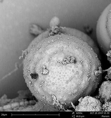
Image © Jan Šťastný
SEM picture of C. pericymatium (var. corrugatum)* in apical view.